Skin biopsies are a common procedure performed by dermatologists to diagnose and evaluate various skin conditions. Whether it’s a suspicious mole, rash, or lesion, a skin biopsy provides valuable insights into the underlying cause and guides appropriate treatment. In this blog post, we’ll explore what a skin biopsy is, why dermatologists recommend it, the different types of skin biopsy, how the skin is analyzed after the procedure, risks and benefits, and post-biopsy care.
What is a Skin Biopsy?
“A skin biopsy is a procedure in which a small sample of skin tissue is removed for examination under a microscope,” explains Dr. Adam Mamelak, Dermatologist and skin cancer expert in Austin, Texas. Dermatologists may recommend a skin biopsy to diagnose or evaluate a wide range of skin conditions, including skin cancer, inflammatory skin disorders, infections, and autoimmune diseases.
Why Might a Dermatologist Recommend a Skin Biopsy?
– Diagnosis: To confirm or rule out a suspected skin condition, such as skin cancer or dermatitis.
– Evaluation: To assess the severity, extent, or progression of a skin condition.
– Treatment Planning: To guide treatment decisions, such as determining the most appropriate medication or therapy.
Types of Skin Biopsy:
1. Punch Biopsy: A small cylindrical tool is used to remove a sample of tissue from the skin.
2. Shave Biopsy: A scalpel or razor blade is used to shave off the top layers of the skin to obtain a sample.
3. Incisional Biopsy: A portion of the lesion is surgically removed for examination.
4. Excisional Biopsy: The entire lesion, along with a margin of normal skin, is surgically removed and examined.
How is a Skin Biopsy Performed?
The specific technique used to perform a skin biopsy may vary depending on factors such as the size, location, and nature of the lesion being sampled. However, the general steps involved in a skin biopsy procedure typically include:
1. Preparation: The dermatologist will clean the area of the skin to be biopsied and may administer a local anesthetic to numb the area and minimize discomfort during the procedure.
2. Biopsy Technique:
- Punch Biopsy: For a punch biopsy, the dermatologist will use a small, circular tool called a punch biopsy instrument to remove a cylindrical sample of skin tissue, including the epidermis, dermis, and superficial subcutaneous tissue.
- Shave Biopsy: In a shave biopsy, the dermatologist will use a sharp blade or razor to shave off the top layers of the skin, obtaining a thin sample of tissue.
- Incisional or Excisional Biopsy: For larger lesions or those suspected of being skin cancer, an incisional or excisional biopsy may be performed. In an incisional biopsy, a portion of the lesion is surgically removed, while in an excisional biopsy, the entire lesion, along with a margin of normal skin, is excised.
3. Hemostasis: After the biopsy sample is obtained, any bleeding from the biopsy site is typically controlled using pressure, cautery, or a topical hemostatic agent.
4. Wound Care: Depending on the type of biopsy performed and the size of the wound, the dermatologist may apply a sterile dressing or adhesive bandage to protect the biopsy site and promote healing.
5. Specimen Handling: The biopsy sample is carefully labeled and placed in a fixative solution to preserve the tissue structure before being sent to a pathology laboratory for processing and analysis.
Overall, a skin biopsy is a relatively quick and straightforward procedure performed in a dermatologist’s office or outpatient setting. While some discomfort or minor bleeding may occur during the biopsy, most patients tolerate the procedure well, and the potential benefits of accurate diagnosis and treatment guidance outweigh any temporary discomfort.
Analyzing Skin Biopsy Samples:
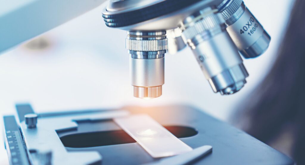
After a skin biopsy, the tissue sample is sent to a pathology laboratory, where it is processed, embedded in paraffin wax, sliced into thin sections, and stained with special dyes. “A pathologist then examines the tissue under a microscope to identify any abnormalities, such as cancer cells, inflammation, or signs of infection,” says Dr. Mamelak.
Risks and Benefits:
Risks:
– Pain or discomfort during the procedure.
– Bleeding, bruising, or infection at the biopsy site.
– Scarring or changes in skin texture.
Benefits:
– Accurate diagnosis of skin conditions.
– Guidance for appropriate treatment.
– Peace of mind for patients and healthcare providers.
Post-Biopsy Care and Healing Expectations:
– Care for the Biopsy Site: Keep the biopsy site clean and dry, and avoid picking at or scratching the area.
– Dressing Changes: Follow any instructions provided by your dermatologist regarding dressing changes and wound care.
– Healing Time: Most skin biopsy sites heal within a few weeks, although healing time may vary depending on the type and location of the biopsy.
Conclusion: Prioritizing Skin Health and Wellness
Skin biopsies are valuable tools in dermatology, providing essential information for diagnosing and treating various skin conditions. By understanding the procedure, risks, benefits, and post-biopsy care, patients can approach the process with confidence and peace of mind. If you have any concerns or questions about a skin biopsy, don’t hesitate to discuss them with your dermatologist—they are there to guide you every step of the way towards optimal skin health and wellness.

